
Dog Anatomy, Animal Anatomy, Anatomy Study, Anatomy Reference, Vet Med, Medical Illustration
It forms the caudal third of the os coxae. Then it enters into the formation of the acetabulum, obturator foramen, and pelvic symphysis. The pubis of a dog is a dorsoventrally compressed bone that extends from the ilium and ischium laterally to the symphysis pubis medially. Dog femur bone The dog femur is the heaviest bone in the hindlimb.

coxae bone DriverLayer Search Engine
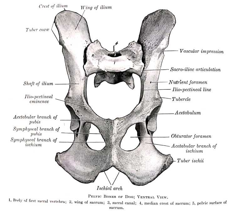
Anatomy. The canine pelvis is composed of the paired os coxae (or hip bone), the sacrum, and the first caudal vertebrae. Each os coxae is developmentally composed of the ilium, ischium, pubis, and acetabular bones, which fuse at 12 weeks of age in the dog. 37 The ilium is divided for practical purposes into the flattened and laterally concave cranial portion known as the ilial wing, and the.

Os coxaelateral view Diagram Quizlet
Max Length: 56-226 mm. Max Proximal Width: -1000 mm. Max Distal Width: -1000 mm. See all OsCoxae samples.

Pelvic Anatomy Canine Human Anatomy
Complete Comparative Anatomy of Horse, Ox and DogVETS GUIDEDVM is a professional program.. Veterinarian can diagnose, treat and manage health issues of la.

BIOL 2325 Lab 1 Os Coxae (Medial and Lateral Views) Diagram Quizlet
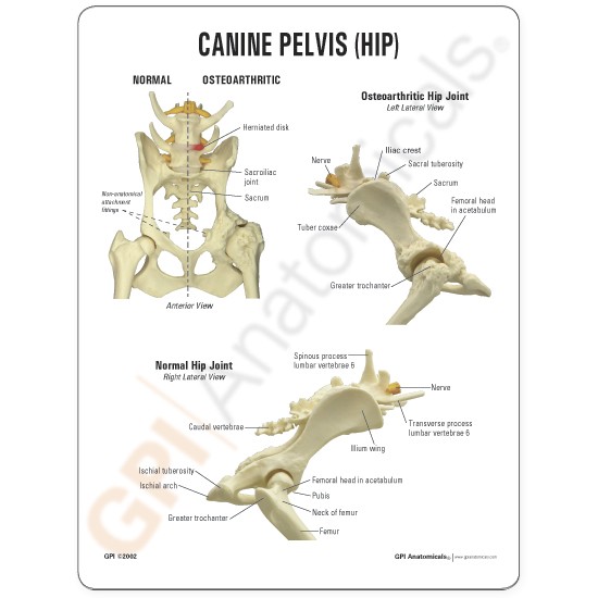
The pelvis is composed of the sacrum and two hip bones (called the os coxae) that unite ventrally at the pelvic symphysis. Sacrum: the sacrum consists of 5 fused sacral vertebrae. The sacroiliac joint(s) are formed by the overlapping of the wing of the sacrum and the wing of the ilium. The sacrum also has (dorsal and ventral) sacral foramina.
Canine Pelvis Hip Anatomical Model
pelvic bones (ossa coxarum) dog 3D Model vetanatMunich pro 13.2k 106 Triangles: 51.2k Vertices: 25.6k More model information canine hip bone, os coxae, innominate bone, pelvic bone or coxal bone linkes und rechtes Hüftbein eine Hundes miteinander verbunden in der Symphysis pelvis Published 4 years ago Animals & pets 3D Models

os coxae dog Diagram Quizlet
25/04/2023 28/01/2022 by Sonnet Poddar The dog pelvis anatomy includes the hip bones, sacrum, muscles, organs, and other associated structures. It is so difficult to explain the detailed anatomical facts of every single part of the dog pelvis in a single article.
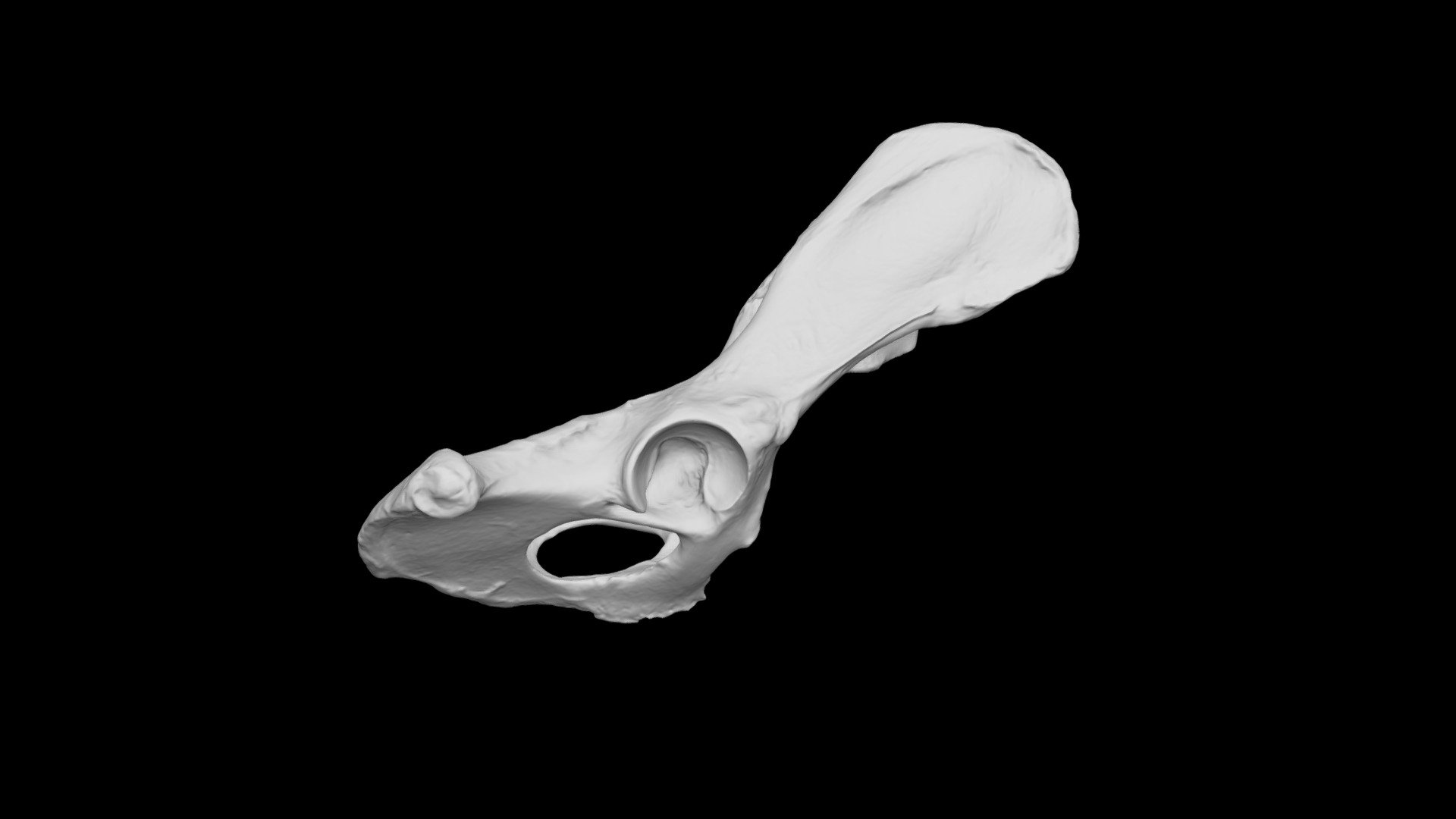
Dog ossa coxae 3D model by Dr. Bobick's Virtual Anatomy Lab (drbobick) [f9f1fcb] Sketchfab
The two hip bones (also called coxal bones or os coxae) are together called the pelvic girdle (hip girdle) and serve as the attachment point for each lower limb. When the two hip bones are combined with the sacrum and coccyx of the axial skeleton, they are referred to as the pelvis.

Anatomy of Os coxae/Pelvic Girdle of Dog with Muscular Attachment Veterinary Anatomy Dog
( Ox, Sheep and Goat, Horse, Pig, Dog, Rabbit, Fowl) Ox The os coxae or hip bone consists of three flat bones, ilium, ischium and pubis, which fuse together to form the acetabulum. The ilium extends from the acetabulum upwards forming the lateral wall of the pelvic cavity.

ventral aspect of canine pelvis (os coxae) Diagram Quizlet
To assist communication among human rehabilitation and veterinary colleagues, some anatomic terms used for dogs appear in regular print with the analogous terminology for humans in parentheses following the canine term. These comparisons have been minimized, as this is a chapter about canine anatomy and not a chapter about comparative anatomy.

Dorsal view of the oscoxae of chinkara showing tuber coxae continue... Download Scientific
The pelvic girdle consists of the ilium, ischium, and pubis bone which is known as the os coxae. As the animal matures, the acetabular bone fuses with the three bones to form the acetabulum.

Dog Os Coxae OsteoID Bone Identification
Os coxae/Pelvic Girdle of Dog with Musuclar Attachment | Veterinary Anatomy | Dog HindlimbAslam u alikmMy name is Dr. Talha Shafiq. So, today I come up with.

Wykres os coxae dog Quizlet
scapula & os coxae(hip bone) Heterotopic bones — os penis [ carnivore; rodent ] os cardis [ cattle ] Shape: Long bones — length greater than diameter. The dog has 321 bones. Regions of a Long Bone Structure of a Long Bone articular cartilage nutrient artery entering nutrient foramen marrow cavity compact bone spongy

Hueso coxal Osteología canina ilustraciones Hueso coxal, Anatomia veterinaria, Anatomía del
HIP BONE (OS COXAE): in young animals each hip bone comprises three bones: ILIUM (OS ILII) - craniodorsal. PUBIS (OS PUBIS) - cranioventral. ISHIUM (OS ISCHI) - caudoventral. all three bones united by a synchondrosis. the synchondrosis ossifies later in life. Hip bones of an ox, left lateral aspect. Hip bones of an ox, ventrocranial aspect.

Pelvic Anatomy Dog Pelvis Anatomy The Institute Of Canine Biology
The pelvis is composed of two hip bones, which are called the os coxae, united ventrally at the pelvic symphysis. Dorsally the two os coxae articulate with each side of the sacrum (at the sacroiliac joints), which are the wings of the sacrum that project ventrally. Each os coxa is formed by the ilium, ischium, pubis, and a small acetabular bone.
Pelvic Girdle Gross Anatomy Anjani Mishra
Hip or os coxae of a dog Osteological features of the canine hip bone Dog acetabulum structure Dog femur head anatomy How is the dog's hip joint formed? Capsular ligament of the dog's hip Cotyloid or transverse ligament of canine hip joint Round ligament of dog hip anatomy Dog hip anatomy muscles Psoas minor and major muscle of the dog hip
